Clinical Cases
MAKE IT VISIBLE.

From fine-detail protocols to motion and metal-artifact management, PreXion Elite combines acquisition, positioning and workflow technologies across a broad clinical range.
CBCT uses a 0.17 mm effective focal spot, supporting fine-detail imaging. PAN/CEPH uses a 0.5 mm focal spot.
A high-resolution acquisition protocol for applications where very fine volumetric sampling is clinically relevant.
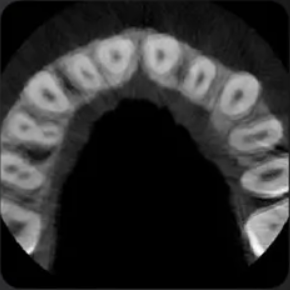
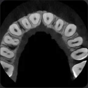
Patient Motion Correction is designed to address motion-related degradation and support image consistency.
Metal Artifact Reduction provides three selectable levels to help manage artifacts associated with high-density materials.
Advanced Position System provides physical stabilization and positioning support during acquisition.
Full-head stitching capability expands visualization options for larger anatomical regions.
CBCT, panoramic and cephalometric imaging within one platform, supporting a broader diagnostic workflow.
The system monitors temperature and energy conditions to support high-throughput operation. PreXion states more than 300 examinations per day without cooling intervals.
Drag the slider to compare the same anatomical region with correction (left) and without correction (right).
Patient Motion Correction
Metal Artifact Reduction
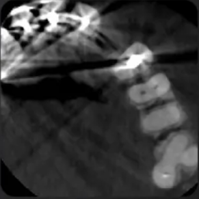
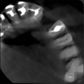
Illustrative comparisons from the supplied radiographic images; original image detail is preserved.
Choose a clinical area to move directly into the relevant field of view and case library.
5×5 · 9×9 · 16×15
9×9 · 16×15
5×5 · 9×9 · 16×15
16×15 · 16×21
Open a case to view the clinical board and its associated video.
Explore panoramic processing filters and lateral cephalometric examinations.
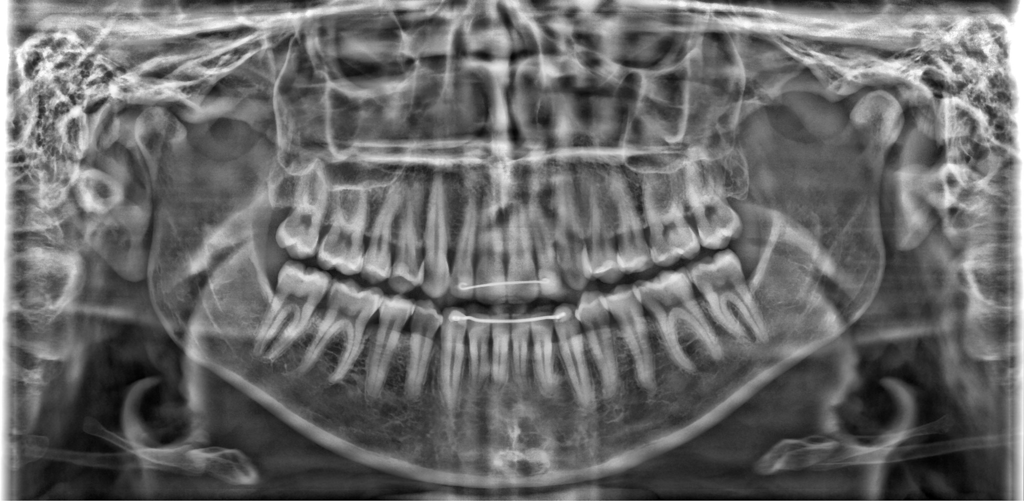
Access clinical image viewers and related datasets when available.
Download packages will be linked here after permanent hosting and authorization checks are complete.
ViewerPrexionElite.zip · 5.46 GB · Opens Google Drive